
How to Make Your Event More Dynamic and Engaging
- April 1, 2026
- lifestyle
Planning an event is one thing, making it truly memorable is another. Whether you’re organising a corporate gathering, private party,… Read More

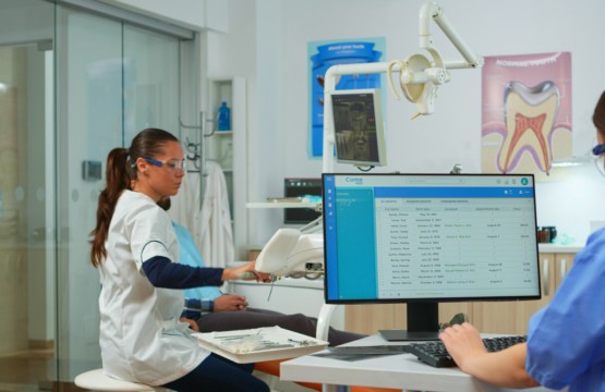
Early and accurate diagnosis is crucial in dentistry, as undetected pathologies can lead to serious complications over time. Traditional radiographic interpretation, while effective, has its limitations—some conditions remain hidden due to the complexity of anatomical structures or subtle early-stage signs. AI-powered solutions like Diagnocat are transforming the way dental professionals analyze imaging, helping uncover pathologies that might otherwise go unnoticed. In this case study, we explore how AI-assisted CBCT interpretation played a vital role in detecting a hidden pathology, leading to timely and effective treatment.
Patient Case Overview
A 42-year-old patient visited a dental clinic complaining of mild but persistent discomfort in the lower left molar region. Standard intraoral X-rays revealed no obvious abnormalities, and a physical examination showed no visible signs of infection or inflammation. Given the patient’s symptoms, the dentist decided to conduct a Cone Beam Computed Tomography (CBCT) scan to get a more comprehensive view.
The Challenge: Traditional Interpretation vs. AI Analysis
Upon reviewing the CBCT scan manually, the dentist initially observed no significant findings that would explain the patient’s symptoms. The scan displayed normal bone structure and no apparent lesions, fractures, or infections. However, to ensure nothing was overlooked, the clinic decided to run the scan through an AI-powered diagnostic tool.
The AI system analyzed the CBCT scan and flagged a small but critical abnormality—a subtle periapical lesion near the roots of a previously treated tooth. The lesion was in its early stages and barely noticeable to the human eye, especially without clear signs of inflammation. Without AI assistance, this pathology might have gone undetected until it worsened, potentially leading to bone loss or a chronic infection.
The Diagnosis and Treatment Plan
With the AI’s findings, the dentist took a closer look at the flagged region, confirming the presence of an early-stage apical periodontitis that had not yet become symptomatic enough to be easily diagnosed. The patient was immediately scheduled for non-surgical retreatment of the affected tooth. By addressing the issue early, the dentist prevented the need for more invasive procedures such as extraction or surgical intervention.
How AI Transformed the Outcome
This case highlights the significant advantages of AI-powered diagnostics in dental imaging:
The Future of AI in Dental Diagnostics
As AI technology continues to evolve, its role in dental imaging will become even more indispensable. AI can assist in detecting complex cases such as cysts, fractures, and hidden infections with remarkable precision. Additionally, AI-powered diagnostics provide standardized assessments, ensuring that all patients receive consistent and reliable evaluations, regardless of the practitioner’s experience level.
By incorporating AI-driven diagnostic tools like Diagnocat, dental professionals can elevate their practice, enhance patient outcomes, and ensure that hidden pathologies do not go unnoticed. This case study is just one example of how AI is revolutionizing dental radiology, making early detection and timely treatment more accessible than ever before.